
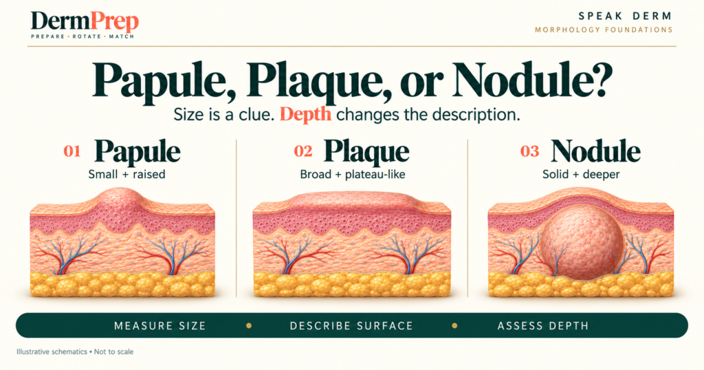
A practical guide to choosing the primary lesion term and explaining your reasoning on rotation.
By the end of this article, you will be able to distinguish papules, plaques, and nodules using measured size, surface contour, and palpable depth—and explain what the term tumor does and does not tell you.
Two lesions can each measure 2 cm and still deserve different descriptions. One may spread across the surface as a plateau; the other may have a substantial component beneath it. The measurement is useful, but it is not the whole examination.
The first Speak Derm article introduced a sequence for describing skin lesions. This follow-up focuses on one decision within that sequence: choosing between papule, plaque, and nodule without treating them as three rungs on a size ladder.
The comparison to keep beside you
| Term | Practical description | What to clarify |
|---|---|---|
| Papule | Small, solid, palpable elevation; commonly less than 1 cm. | Measured diameter and surface shape. |
| Plaque | Broad, palpable area, usually greater than 1 cm and often plateau-like. | Contour and any scale or other surface change. |
| Nodule | A lesion with a deeper dermal or subcutaneous component. | Depth and consistency, rather than diameter alone. |
| Tumor | An abnormal tissue mass that may be benign or malignant. | Describe the examination separately from the diagnosis. |
Definitions summarized from clinical terminology resources and the National Cancer Institute. [1,2,4] This comparison supports description; it is not a diagnostic algorithm.
A papule does not have to be dome shaped
Papules may be pointed, flat-topped, umbilicated, or rough rather than rounded. Shape modifies the description; it does not replace the primary lesion term. A small, flat-topped elevation can therefore still be a papule. [1]
In a teaching description, “a 4 mm flat-topped papule” tells the listener more than “a small bump.” Add color, site, and surface findings only when you have actually observed them.
A plaque is broad but need not be scaly
A plateau-like surface is a useful starting image, but plaques vary in contour and may be thickened without obvious elevation. Some develop through coalescence of papules. Scale is a separate surface finding, not a requirement for calling a lesion a plaque. [1,2]
Consider the phrase “a 2 cm plaque with overlying scale.” The noun identifies the primary morphology; the final phrase adds surface information. Calling it simply “scale” loses the underlying lesion.
A nodule asks you to assess depth
Nodules extend into the dermis or subcutaneous tissue. Their visible surface may not represent their full extent. Inspection and palpation answer different questions: you can see contour, but examination adds information about the component beneath it. [1,2,3]
Do not teach this as “anything larger than 1 cm is a nodule.” A broad plaque may exceed that measurement. Instead, explain the deeper component that supports your choice.
A clinical examination can suggest depth; it does not supply a microscopic map. If you cannot confidently localize the lesion, describe a palpable deeper component rather than asserting an exact tissue layer.
Use measurements when definitions differ
DermNet includes lesions measuring exactly 1 cm in its papule definition, while Stanford uses less than 1 cm and Merck describes papules as usually less than 10 mm. These wording differences are a reason to document the actual size, not to omit the term. [1,2,3]
At a boundary, say what you measured and describe the architecture. “A 10 mm solid elevation without an appreciable deeper component” communicates the finding even when a teaching convention differs. Follow your service’s terminology while retaining the measurement.
What tumor means in this discussion
The National Cancer Institute defines a tumor as an abnormal tissue mass and distinguishes benign from malignant tumors. The word alone does not establish cancer. [4] It is therefore unhelpful to treat “tumor” as a fourth size category that automatically follows papule, plaque, and nodule.
Keep your description and interpretation separate. Document the lesion’s size, contour, consistency, and surface, then state the diagnostic concern and what remains unknown. A reassuring-sounding morphology term cannot substitute for clinical assessment, and a concerning-sounding term cannot substitute for a diagnosis.
Turn uncertainty into a useful presentation
Start with location and measured size, name the best-supported primary morphology, and add the finding that justifies it. Accurate description is the foundation of the dermatologic examination. [3]
The following are hypothetical teaching examples, not patient encounters:
- Papule: “On the forearm is a solitary 5 mm smooth, skin-colored papule.”
- Plaque: “On the elbow is a 2 cm sharply demarcated plaque with overlying scale.”
- Nodule: “On the upper arm is a 1.5 cm firm nodule with a palpable deeper component.”
- Uncertain depth: “The photograph shows a raised lesion, but I cannot determine its depth without palpation.”
The last statement identifies missing examination data rather than guessing. Do not add tenderness, mobility, fluctuance, or negative findings unless they were assessed.
Try three questions
These hypothetical examples test description, not diagnosis. Answer before reading the explanations.
- A solid, flat-topped elevation measures 4 mm. Does the flat top rule out a papule?
- A 2.5 cm broad, raised lesion has surface scale. What primary morphology and secondary finding would you report?
- A 1.5 cm firm lesion has a palpable deeper component. What term fits, and does that term establish malignancy?
Answers and explanations
- No. Papules can be flat-topped. Report the measured size and contour together. [1]
- A plaque with scale. The broad architecture supports plaque; scale is reported separately. [2]
- A nodule. The deeper component supports the term. It is a morphology description, not a determination of benign or malignant behavior. [2,4]
The takeaway for your next presentation
Measure the lesion, describe its surface, and assess its depth. When the term is uncertain, keep the observations precise. The goal is a description another clinician can understand and reassess.
References
- Oakley A. Terminology in dermatology. DermNet.
- Merck Manual Professional Edition. Description of skin lesions.
- Stanford Medicine 25. The general dermatology exam: learning the language.
- National Cancer Institute. Tumor. NCI Dictionary of Cancer Terms.
Sources accessed September 16, 2026.
Correction or editorial concern: hello@dermprep.com
Medical disclaimer: DermPrep is an independent educational platform. Content is provided for educational purposes only and is not a substitute for professional medical advice, diagnosis, treatment, or independent clinical judgment.