
Key Takeaways
- A diagnosis should follow observation. Begin with what is visible and palpable rather than the disease name.
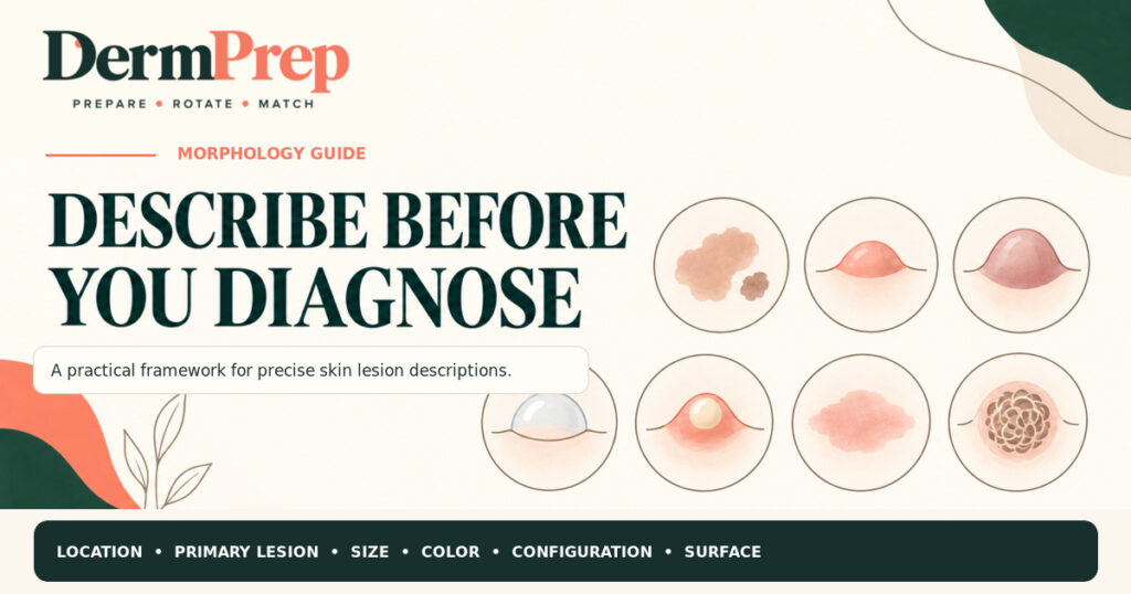
- Use the same sequence every time: location and distribution, primary lesion, size, color, configuration, border, and surface or secondary change.
- Measure lesions instead of relying only on labels such as small or large; size thresholds for some morphology terms vary among references.
- Inflammation does not always look bright red. Describe the hue you actually see and use palpation, texture, scale, warmth, edema, and symptoms as additional clues.
- A strong presentation allows another clinician to picture the eruption before you give the differential diagnosis.
Why Rash Is Not Enough
“Rash” may identify the reason for the visit, but it does not tell another clinician what is on the skin. A useful dermatologic description should let the listener build a mental image without seeing the patient. That requires a shared vocabulary for morphology, configuration, and distribution. Standardized terminology also forces the examiner to slow down and notice findings that may narrow the differential diagnosis.[1–3]
The practical rule is simple: describe before you diagnose. If your first thought is “psoriasis,” pause. What can you defend from the examination? Perhaps the patient has multiple, symmetric, sharply demarcated plaques with overlying scale on the extensor elbows and knees. That description carries clinical information; the diagnostic label comes next.
The DermPrep Description Sequence
Use the same order for every patient until the sequence becomes automatic:
- Location and distribution. Where is it, and how is it distributed? Note the precise sites, localized versus generalized involvement, symmetry, and patterns such as acral, flexural, extensor, intertriginous, dermatomal, photodistributed, or mucosal.
- Primary lesion. Identify the basic lesion that best represents the process before scratching, crusting, erosion, treatment, or infection altered it.
- Number and size. State whether the lesions are solitary, few, multiple, or numerous. Measure a representative lesion in millimeters or centimeters.
- Color. Name the hue you actually see: pink, red, violaceous, brown, gray, white, yellow, blue, black, hyperpigmented, or hypopigmented. Avoid using erythematous as an automatic substitute for observation.
- Shape and configuration. Describe the shape of individual lesions and how multiple lesions relate to one another. Examples include round, oval, annular, arcuate, linear, grouped, confluent, reticulated, targetoid, serpiginous, or herpetiform.
- Border and surface. Note whether the border is sharply or poorly demarcated. Then describe scale, crust, erosion, ulceration, excoriation, fissuring, lichenification, atrophy, scarring, or other secondary change.
- Palpation and associated findings. When appropriate, assess temperature, tenderness, induration, fluctuance, mobility, blanching, and depth. Add relevant symptoms and findings involving hair, nails, or mucosa.
Start With the Primary Lesion
Primary morphology describes the lesion produced by the underlying process. Secondary morphology describes changes that develop afterward, whether from disease evolution, scratching, infection, trauma, or treatment. Look for the least altered representative lesion, including at the edge of an eruption when the center has been excoriated or eroded.
| Primary lesion | What to look for |
| Macule or patch | Flat change in color without elevation or depression. A patch is larger than a macule. |
| Papule or plaque | Solid, elevated lesion. A plaque is broader and often formed by enlargement or coalescence of papules. |
| Nodule | Solid lesion with greater depth in the dermis or subcutis; palpation helps establish depth. |
| Vesicle or bulla | Fluid-filled blister. A bulla is larger than a vesicle. |
| Pustule | Circumscribed lesion containing purulent material; the content may be sterile or infectious. |
| Wheal | Transient, edematous, elevated lesion, classically associated with urticaria. |
| Cyst | Encapsulated or circumscribed cavity containing fluid or semisolid material. |
A note about size: Published references do not use perfectly uniform cutoffs for every lesion term. Many teaching resources use 1 cm to distinguish macule from patch, papule from plaque, and vesicle from bulla, while other sources use different thresholds.[1–3] Measure the lesion and describe its depth and contents; those observations are more portable than relying on a cutoff alone.
Secondary Change Is Not the Primary Lesion
- Scale: Visible accumulation or shedding of stratum corneum.
- Crust: Dried serum, blood, or purulent exudate on the surface.
- Erosion: Superficial loss of epidermis; generally heals without scarring.
- Ulcer: Loss of epidermis and at least part of the dermis; may heal with scarring.
- Excoriation: Linear or punctate erosion caused by scratching or manipulation.
- Fissure: Linear crack extending into the epidermis and sometimes dermis.
- Lichenification: Thickening with accentuated skin markings from chronic rubbing or scratching.
- Atrophy or scar: Thinning of tissue or fibrous replacement following injury or inflammation.
Describe Color Across Skin Tones
Color is an observation, not a fixed property of a diagnosis. Inflammatory change may appear pink or red, but in more deeply pigmented skin it may be subtler or appear violaceous, brown, gray, or as a change from the patient’s baseline tone. Reliance on visible redness alone can underestimate inflammation.[4]
Use adequate lighting and compare involved skin with nearby uninvolved skin when possible. Name the visible hue, then add other evidence of activity: warmth, edema, tenderness, induration, scale, texture, and symptoms such as itch or pain. Palms, soles, mucosa, and nail beds may offer additional contrast, but they should not replace examination of the affected site.
Avoid assuming that every dark area represents active inflammation. Postinflammatory pigment alteration may remain after an inflammatory eruption has improved. The rest of the examination and the time course help separate residual pigment from active disease.
Say It Like This
Use this presentation formula:
On the [location], there is/are [number] [color] [primary lesion or lesions] measuring [size], with [shape or configuration], [border], and [surface or secondary change], distributed [pattern]. Associated findings include [relevant palpation, mucosal, hair, nail, or symptom findings].
Example: On the extensor elbows and knees, there are multiple symmetric, sharply demarcated erythematous-to-violaceous plaques with overlying micaceous scale. No mucosal involvement is present.
The description supports a focused differential, but it does not pretend that morphology alone proves the diagnosis. History, full examination, dermoscopy, testing, biopsy, and clinical context may still be necessary.
From Description to Differential
After the description, ask which diagnoses best explain the combination of morphology and distribution. Rank rather than list:
- Most likely: the diagnosis that best fits the complete pattern.
- Important mimic: a plausible alternative with an overlapping appearance.
- Must not miss: a less common diagnosis whose consequences require timely recognition.
- Discriminator: the history, examination finding, bedside test, laboratory study, dermoscopy, or biopsy result that would change the ranking.
Rotation Ready Pearls
- Lead with morphology, not “the patient has a rash” or an unqualified diagnosis.
- Measure at least one representative lesion and say whether the process is superficial or deep, solid or fluid filled.
- Search for an early or less manipulated lesion when scratching, crust, or erosion obscures the primary morphology.
- Distribution often carries as much diagnostic weight as the individual lesion. Mention palms, soles, scalp, mucosa, nails, and intertriginous sites when relevant.
- Describe the color you see across the patient’s baseline skin tone; do not make bright redness a requirement for inflammation.
- Finish with a ranked differential and the feature you would use to separate the leading possibilities.
Common Pitfalls
Diagnosing before describing. A label can anchor your examination and make you overlook contradictory findings.
Calling the secondary change the primary lesion. Scale, crust, and excoriation may hide the original macule, papule, plaque, vesicle, or nodule.
Ignoring distribution. A “scaly plaque” remains broad; extensor, flexural, annular, dermatomal, or photodistributed patterns narrow the problem.
Using erythematous automatically. If the lesion is violaceous, brown, gray, or only subtly different from baseline, say so.
Listing everything. A differential becomes useful when it is prioritized and linked to discriminating findings.
Before You Go
Try these without looking back at the framework.
- A patient has several sharply demarcated, solid, elevated lesions measuring 2–3 cm with overlying scale. Which primary lesion term is most appropriate?
- Why should you look for a less excoriated lesion before assigning the primary morphology?
- Inflammation is suspected, but the involved skin appears violaceous rather than bright red. What should you document and assess next?
Answers and Explanations
1. Plaques. They are broad, solid, elevated lesions. Scale is a secondary surface change and should be described separately.
2. The primary lesion may be obscured. Scratching can create excoriation, erosion, crust, or lichenification. A less altered lesion better reflects the underlying process.
3. Describe the actual hue and use additional signs of activity. Document violaceous color relative to baseline and assess warmth, edema, tenderness, induration, scale, texture, symptoms, distribution, and relevant mucosal or appendageal findings. Bright redness is not required for inflammation.
The One Sentence to Remember
Before naming the diagnosis, describe the location and distribution, primary lesion, size, color, configuration, border, and surface. Then rank the differential and explain what would separate the leading possibilities.
Save this framework for your next dermatology rotation, then practice applying it to unfamiliar images in Pimp Me.
References
- Nast A, Griffiths CEM, Hay R, Sterry W, Bolognia JL. The 2016 International League of Dermatological Societies’ revised glossary for the description of cutaneous lesions. Br J Dermatol. 2016;174(6):1351–1358. doi:10.1111/bjd.14419. Source
- Stanford Medicine 25. The General Dermatology Exam: Learning the Language. Accessed September 15, 2026. Source
- Merck Manual Professional Edition. Description of Skin Lesions. Accessed September 15, 2026. Source
- Forsyth A, Prajapati S, Frasier KM, et al. Diagnostic Disparities in Erythema Visibility: A Call to Redefine Inflammatory Assessment in Diverse Skin Tones. Cureus. 2025;17(10):e94930. doi:10.7759/cureus.94930. Source
- Rimoin L, Altieri L, Craft N, Krasne S, Kellman PJ. Training pattern recognition of skin lesion morphology, configuration, and distribution. J Am Acad Dermatol. 2015;72(3):489–495. doi:10.1016/j.jaad.2014.11.016. Source
Correction or editorial concern: hello@dermprep.com
Medical disclaimer: DermPrep is an independent educational platform. Content is provided for educational purposes only and is not a substitute for professional medical advice, diagnosis, treatment, or independent clinical judgment.